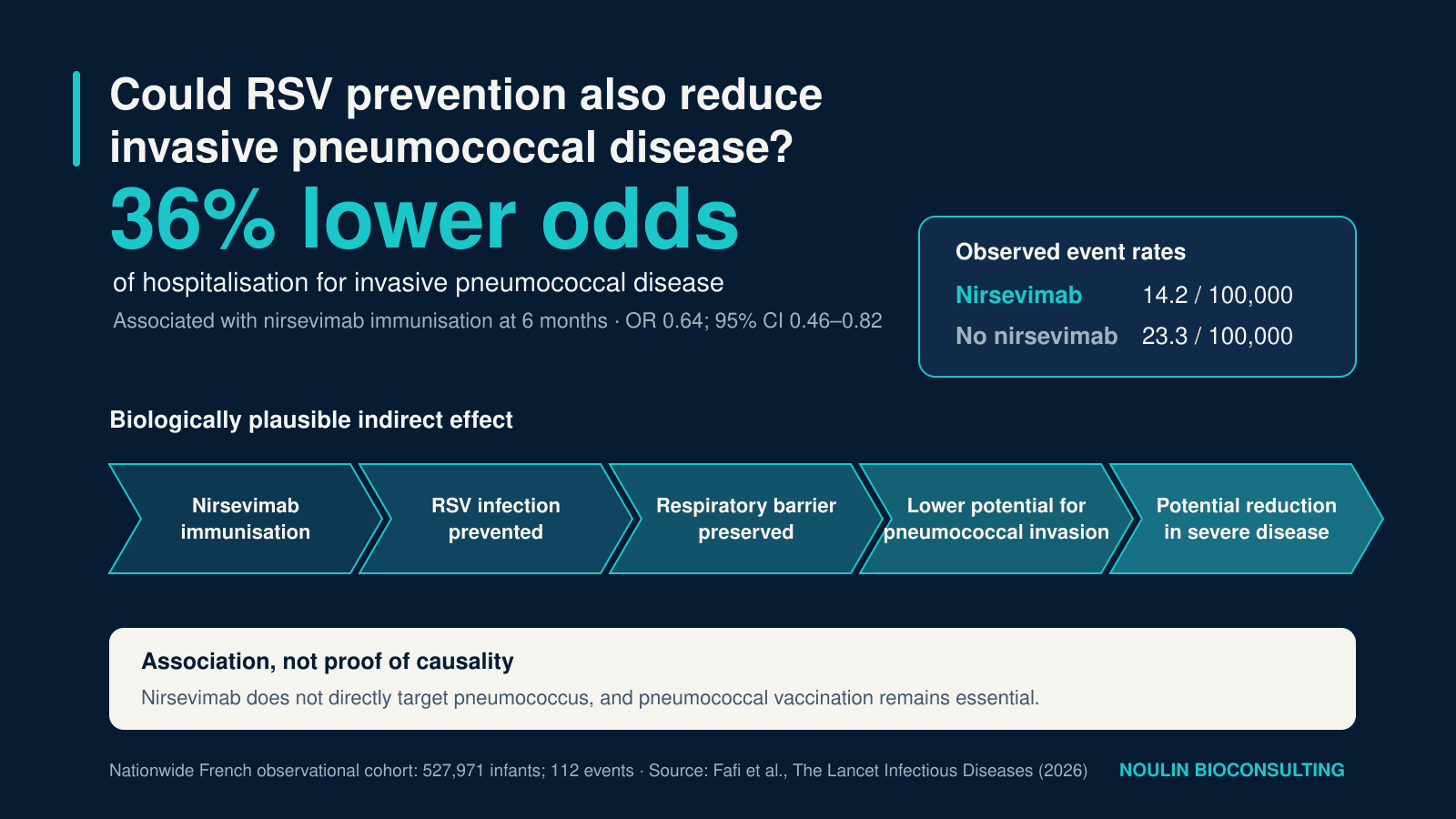
As the French National Authority for Health upgrades its assessment of nirsevimab, a nationwide French study associates nirsevimab with fewer hospitalisations for invasive pneumococcal disease in infants. The finding is consistent with the biology of respiratory coinfections—but it requires confirmation before any causal effect can be claimed.
This summer, two developments brought nirsevimab back to the centre of paediatric prevention in France.
On 15 July 2026, the Transparency Committee of the French National Authority for Health (HAS) upgraded the clinical benefit of Beyfortus from “moderate” to “substantial” [1]. Reimbursement through community pharmacies increased from 30% to 65% on 31 August 2026. The decision, published in the French Official Journal on 21 August, sets the patient contribution at 35% [9].
Just a few days later, a nationwide study published in The Lancet Infectious Diseases suggested that the benefits of nirsevimab may extend beyond the prevention of respiratory syncytial virus (RSV) disease. The authors observed fewer hospitalisations for invasive pneumococcal disease among immunised infants [2].
These developments address distinct questions: the assessment of RSV prevention and a possible indirect bacterial benefit. The pneumococcal finding does not extend the authorised indication.
A Long-Acting Antibody Against RSV
Beyfortus contains nirsevimab, a long-acting monoclonal antibody directed against the RSV fusion protein. Administered directly to the infant, it provides passive protection during the first RSV season [10].
It is therefore neither a paediatric vaccine—because it does not require the infant’s immune system to generate its own antibodies—nor an antibacterial treatment. Nirsevimab has no direct activity against Streptococcus pneumoniae.
If an effect on pneumococcal disease does exist, it would be indirect. By preventing or attenuating RSV infection, nirsevimab could interrupt the sequence of events that sometimes enables pneumococcus to progress from nasopharyngeal colonisation to severe disease.
What the French Study Found
Researchers used the French National Health Data System (SNDS) to establish a retrospective cohort of children born in metropolitan France between 6 February 2023 and 31 January 2024.
Of 608,641 births, 527,971 infants were included in the analysis. A total of 119,435—22.6%—had received nirsevimab.
During six months of follow-up, 112 invasive pneumococcal disease events were identified:
17 among immunised infants, or 14.2 cases per 100,000;
95 among non-immunised infants, or 23.3 cases per 100,000.
After propensity-score weighting to reduce measurable differences between the groups, nirsevimab immunisation was associated with 36% lower odds of hospitalisation for invasive pneumococcal disease (OR 0.64; 95% CI 0.46–0.82). At nine months, the association remained similar, with an estimated reduction of 34% [2].
The relative effect is notable, but the absolute difference also matters: the crude difference between groups was approximately nine fewer invasive infections per 100,000 infants followed for six months. The outcome is fortunately rare, but can be extremely serious when it occurs, including bacteraemia, sepsis and meningitis.
The crude difference is descriptive, not an adjusted estimate of cases prevented. Odds compare the probability of an event with the probability of no event. For a rare outcome, an odds ratio approximates a risk ratio but is not identical to it.
Why Might RSV Promote Pneumococcal Disease?
This signal does not arise in a biological vacuum. A 2024 systematic review concluded that RSV and pneumococcus interact, supported by epidemiological, clinical and mechanistic evidence [3].
Several complementary mechanisms may be involved.
1. A More Permissive Respiratory Barrier
RSV primarily infects ciliated epithelial cells in the respiratory tract. Infection impairs the integrity and function of this first line of defence, disrupts mucociliary clearance and alters the expression of cellular receptors to which respiratory bacteria can adhere.
Cellular models have shown that prior RSV infection increases the adhesion of S. pneumoniae to the respiratory epithelium [5]. This provides a plausible mechanism for denser colonisation or easier crossing of the mucosal barrier.
2. Direct Virus–Bacterium Interaction
Experimental studies have also shown that the RSV G glycoprotein can bind pneumococcal penicillin-binding protein 1a. In these models, the interaction alters bacterial gene expression, increases pneumolysin expression and enhances pneumococcal virulence [6].
These findings strengthen biological plausibility, but they derive mainly from cellular and animal models. On their own, they do not establish that this mechanism accounts for the invasive infections observed in infants.
3. Effects on Immunity and the Respiratory Ecosystem
A viral infection can temporarily alter innate immune responses, local inflammation and the respiratory microbiota. In an infant already colonised with pneumococcus, this environment could promote bacterial proliferation, migration into the lower respiratory tract or invasion of normally sterile sites.
At population level, RSV epidemics and pneumococcal infections also display closely related seasonal patterns. A US analysis covering more than 700,000 RSV hospitalisations estimated that approximately 20% of pneumococcal pneumonia in children younger than one year was statistically attributable to RSV activity [4]. Temporal correlation cannot establish the causal sequence in each child, but it adds to the overall coherence of the hypothesis.
What the Study Does Not Yet Establish
The cohort size and nationwide coverage are major strengths. SNDS data make it possible to investigate a rare outcome under real-world conditions, at a scale that would be difficult to achieve in a conventional clinical trial.
However, the study remains observational.
Residual confounding may persist despite statistical weighting. Families who accept nirsevimab may differ from others in healthcare-seeking behaviour, uptake of other vaccines, housing conditions, childcare arrangements or exposure to tobacco smoke. Not all of these factors are captured accurately in administrative healthcare databases.
In addition:
only 112 invasive infections were observed;
the study does not show that each pneumococcal infection was preceded by laboratory-confirmed RSV infection;
it documents an association between immunisation and hospitalisation, not antibacterial activity of nirsevimab;
the findings come from a single first French immunisation campaign and need to be reproduced across additional seasons and healthcare systems.
The scientifically accurate conclusion is therefore that nirsevimab was associated with fewer invasive pneumococcal infections. Claiming today that it “protects against pneumococcus” would go beyond the evidence.
From Association to Causality: The Role of Human Experimental Models
This question illustrates the complementarity of population-level data and mechanistic studies.
The SNDS can detect a rare clinical signal at national scale, but it cannot fully describe the biological sequence that leads to it. Conversely, a controlled human infection model can closely examine timing, colonisation, mucosal responses and pathogen interactions—without attempting to reproduce invasive disease.
A multicentre protocol published in 2025 was designed specifically to administer RSV-A and serotype 6B pneumococcus sequentially to adult volunteers in a randomised order [7]. Its primary objective is to determine whether prior RSV infection increases the risk of subsequent pneumococcal colonisation. Results from this type of study could help connect epidemiological observations with the early mechanisms of RSV–pneumococcus interaction.
What Could This Mean for Prevention?
In the short term, this study does not change the indication for Beyfortus and clearly does not replace routine pneumococcal vaccination in infants.
It does, however, open several lines of investigation:
include bacterial complications, antibiotic use and all-cause respiratory infections more systematically among the indirect outcomes assessed in RSV prevention programmes;
monitor pneumococcal serotypes and document viral infections preceding invasive cases;
reproduce the analysis across subsequent French campaigns and in other countries;
assess whether this potential benefit changes the health-economic value of broad RSV immunisation;
examine the combined effects of RSV and pneumococcal prevention strategies rather than evaluating them in isolation.
Randomised trials of nirsevimab had already shown reductions in all-cause respiratory infections and antibiotic prescriptions [8]. The new French study provides a more specific signal: preventing a viral infection may also reduce a rare and serious bacterial complication.
A Broader Perspective
The HAS reassessment of Beyfortus reflects the growing strength of real-world evidence for preventing severe RSV disease. The pneumococcal study raises a broader question: how much of the benefit of antiviral prevention lies beyond the disease directly targeted?
The result is biologically plausible and sufficiently compelling to justify further investigation. It is not yet sufficient to support an antipneumococcal indication for nirsevimab.
It nevertheless reinforces a central principle of respiratory infection biology: pathogens do not act in isolation. Preventing the first event in an infectious cascade may produce benefits that only become visible at population scale.
References
Haute Autorité de santé. BEYFORTUS (nirsevimab)—reassessment of clinical benefit and added clinical value, opinion of 15 July 2026.
Fafi I, Valtuille Z, Kaguelidou F, et al. Effectiveness of nirsevimab immunisation on invasive pneumococcal disease in children nationwide in France: an observational cohort study. The Lancet Infectious Diseases. 2026.
Besteman S, Bogaert D, Bont L, et al. Interactions between respiratory syncytial virus and Streptococcus pneumoniae in the pathogenesis of childhood respiratory infections: a systematic review. The Lancet Respiratory Medicine. 2024.
Weinberger DM, Klugman KP, Steiner CA, et al. Association between Respiratory Syncytial Virus Activity and Pneumococcal Disease in Infants: A Time Series Analysis of US Hospitalization Data. PLOS Medicine. 2015.
Avadhanula V, Rodriguez CA, DeVincenzo JP, et al. Respiratory Viruses Augment the Adhesion of Bacterial Pathogens to Respiratory Epithelium in a Viral Species- and Cell Type-Dependent Manner. Journal of Virology. 2006.
Smith CM, Sandrini S, Datta S, et al. Respiratory syncytial virus increases the virulence of Streptococcus pneumoniae by binding to penicillin binding protein 1a. American Journal of Respiratory and Critical Care Medicine. 2014.
Brito-Mutunayagam S, Hamilton DO, Mitsi E, et al. Understanding the interaction of upper respiratory tract infection with respiratory syncytial virus and Streptococcus pneumoniae using a human challenge model: a multicenter, randomized controlled study protocol. PLOS One. 2025.
Simões EAF, Madhi SA, Muller WJ, et al. Efficacy of nirsevimab against respiratory syncytial virus lower respiratory tract infections in preterm and term infants: a pooled analysis of randomised controlled trials. The Lancet Child & Adolescent Health. 2023.
UNCAM. Decision of 4 August 2026, published 21 August 2026, effective 31 August 2026.
European Medicines Agency. Beyfortus: indication and mechanism.
Planning a vaccine, antiviral or human challenge program?
Get independent scientific input on model selection, protocol strategy, endpoints, evidence generation and decision-making.
Scientific brief